

Newsletter Subscribe
Enter your email address below and subscribe to our newsletter

Ideas Worth Building
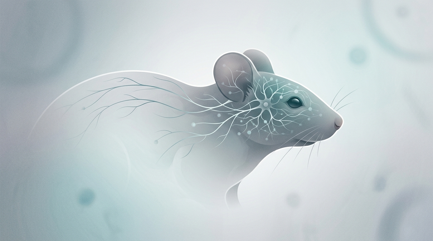
Scientists studying human brain tissue in mice have reported a striking result: transplanted human neural cells survived in an animal nervous system, formed connections, extended toward the spinal cord, and responded when nearby tissue was injured.
The work is not a medical treatment, and it does not mean researchers created “humanlike” mice. But it does add to a growing body of neuroscience research showing that human cells can be studied in living animal models in ways that may reveal how neural circuits develop, connect, and react to damage.
For researchers working on brain development, neurological disease modeling, spinal cord injury, and regenerative medicine, that combination is important. Cells grown in a dish can reveal a great deal. Cells placed into a living nervous system may show how human neural tissue behaves when exposed to blood supply, immune signals, electrical activity, and injury responses that are difficult to reproduce in laboratory culture.
In broad terms, the study involved transplanting human brain tissue or human neural cells into mice and then observing whether those cells could survive and integrate with the host nervous system.
This kind of experiment sits at the intersection of organoid research and transplantation biology. Human brain organoids are small, simplified clusters of neural tissue grown from stem cells. They are not miniature brains, but they can contain multiple types of developing brain cells arranged in ways that help scientists study early human brain development and disease.
One major limitation of organoids grown in laboratory dishes is that they do not fully replicate the environment of a living brain. They may lack mature blood supply, long-range connections, and the complex signaling found inside an organism. Transplantation into a mouse model gives researchers a way to test whether human cells can mature further, receive inputs, send outputs, and participate in living neural circuits.
According to the reported findings, the transplanted human cells did more than simply survive. They formed neural circuits, extended connections in the direction of the spinal cord, and showed a response when injury occurred.
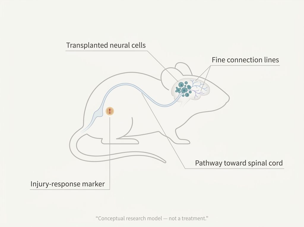
A neural circuit is a network of nerve cells that communicate with one another. In the brain and spinal cord, neurons pass signals through specialized connections called synapses. These networks allow nervous systems to process sensory information, generate movement, regulate behavior, and respond to the body’s needs.
When researchers say transplanted human cells formed circuits, they generally mean the cells developed functional connections with other neurons. That does not imply the transplanted tissue became a complete human brain region or gave the mouse human cognition. It means the cells appeared capable of wiring into a biological network.
That distinction matters. The scientific significance is about integration: whether human neural tissue can connect, signal, and participate in the surrounding nervous system. Integration is a central question for organoid research and regenerative medicine, because future disease models and repair strategies depend on understanding how transplanted cells behave inside living tissue.
One especially notable part of the reported research is that the human cells extended connections toward the spinal cord.
The spinal cord is the body’s main communication pathway between the brain and much of the peripheral nervous system. For scientists studying movement, paralysis, and spinal cord injury, long-distance connections are a major challenge. Neural cells may survive after transplantation, but survival alone is not enough. To be useful in research models, cells must also interact with existing circuits in meaningful ways.
Connections extending toward the spinal cord suggest that transplanted human neural tissue can respond to guidance signals in the host animal and project beyond the immediate transplant site. That could help researchers study how human neurons grow axons, navigate tissue environments, and communicate with motor pathways.
This does not mean a therapy for spinal cord injury is available or imminent. Translating findings from mice to humans is difficult, and the safety requirements for any future human application would be far higher. But as a laboratory model, the observation gives scientists another way to ask how human neural cells might behave after injury or transplantation.
The reported injury response may be just as important as the circuit formation.
In the nervous system, injury triggers a cascade of events. Cells may activate repair programs, inflammatory signals may change, and nearby neurons and support cells may alter their behavior. A transplanted human neural cell that responds to injury inside a mouse model gives researchers a chance to study these processes in a living environment.
That could be useful for understanding neurological disorders, traumatic injury, and the limits of neural repair. Many brain and spinal cord conditions involve not just cell death, but also disrupted communication between cells and abnormal responses to damage.
If human cells in an animal model react in measurable ways, researchers can begin asking more precise questions: Which signals activate the response? Do different types of human cells react differently? Does the host environment help or hinder repair? Could disease-specific human cells behave differently from healthy cells?
Those are research questions, not clinical claims. The current value is in modeling and discovery.
Mouse models remain central to neuroscience because they allow researchers to study living systems in a controlled way. Mice are not humans, and findings in mice often fail to translate directly to people. Still, animal models can reveal processes that are impossible to observe fully in cell culture.
For human brain tissue in mice, the goal is usually not to “humanize” an animal. The goal is to create a controlled system in which human cells can be observed inside a functioning nervous system.
That can help with several areas of research:
– Brain development: Scientists can examine how human neurons mature and connect.
– Disease modeling: Cells carrying disease-related traits may reveal how disorders affect circuits.
– Organoid research: Transplantation may show whether organoid-derived tissue can integrate in vivo.
– Injury biology: Researchers can study how human cells respond to damage and repair signals.
– Regenerative medicine: Early findings can clarify what transplanted cells would need to do before any future therapy is considered.
The key word is “early.” These models are tools for investigation, not evidence that brain repair treatments are ready for patients.
Research involving human neural tissue in animals raises ethical questions that scientists, institutions, and regulators must take seriously.
The main concerns include animal welfare, the degree of human neural cell integration, and whether such experiments could meaningfully alter an animal’s cognition or experience. Current studies are typically designed with limits, oversight, and careful monitoring, but the ethical discussion becomes more important as human neural models become more sophisticated.
Clear language is part of that responsibility. Saying scientists “grew a human brain in a mouse” can mislead the public and exaggerate what happened. A more accurate description is that human neural tissue or cells were transplanted into mice and showed signs of integration, circuit formation, long-range growth, and injury response.
That is still scientifically significant. It just needs to be understood in the right context.
The reported study suggests that transplanted human neural tissue can survive inside mice, form neural circuits, extend toward the spinal cord, and respond to injury. For neuroscience, that could improve how researchers model human brain development, neurological disease, and nervous system repair.
But the finding is not a clinical breakthrough and should not be read as a near-term treatment for brain or spinal cord injury. It is an early-stage animal-model study that may help scientists ask better questions about how human cells connect, communicate, and react inside living nervous systems.
Read the Research: For full experimental details, methods, limitations, and author conclusions, consult the original peer-reviewed study and any accompanying journal or institutional materials.